X-Ray in Ooty
Dental X-rays show what an examination cannot: decay between teeth, bone levels around roots, root canal anatomy, impacted teeth and hidden infection. At RC's Dentcare in Ooty, radiographs are taken only when they will change diagnosis or treatment, and the images are reviewed on screen with you.
Half of every tooth is out of sight
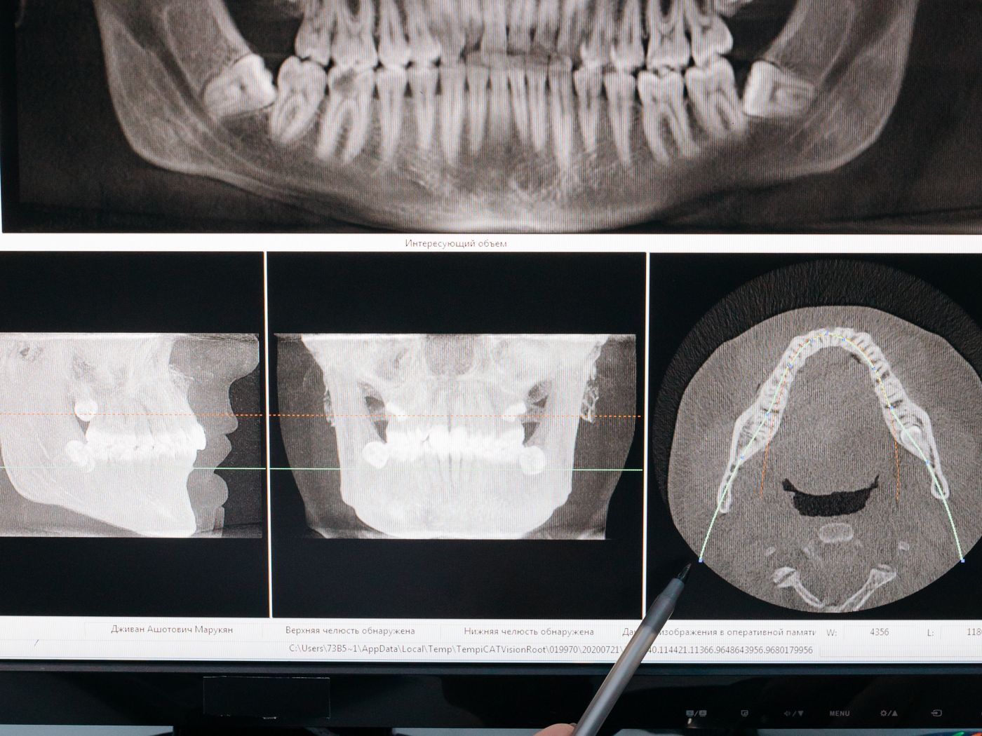
A visual examination can assess the biting surfaces and the visible outer faces of your teeth. It cannot see between two teeth that are touching, and it cannot see anything below the gum — which is where roots, bone and abscesses are.
That is not a small blind spot. It is where a large share of dental disease begins.
Diagnosing without imaging means guessing
Treating a painful tooth without a radiograph risks treating the wrong tooth, missing a second canal, or restoring a tooth with a vertical root fracture that was never going to be saved. Placing an implant without imaging bone height is not a plan, it is a hope.
The counterweight is that imaging should not be routine or automatic. Every exposure should have a reason, which is why we justify each one against what it will actually change.
How we use imaging here
We request the specific view that answers the clinical question, capture it with protective protocols in place, then interpret it with you on the screen so you can see what we are describing rather than take it on trust.
Images are kept so that future radiographs can be compared against them — change over time is often more informative than any single picture.
If you are unsure whether a problem needs imaging, an examination will settle it before anything is taken.
Book a dental examination with imaging
What to expect
- Justified, need-based imaging
- Protective protocols followed
- Images explained on screen with you
- Records kept for comparison over time
Your treatment journey
Step 01
Justify
Imaging requested only when it informs treatment.
Step 02
Capture
The appropriate view is taken.
Step 03
Interpret
Findings reviewed with you.
Step 04
Plan
Treatment adjusted to what the image shows.
X-Ray in Ooty — common questions
- Are dental X-rays safe?
- Modern digital dental radiography uses a very low dose, and protective protocols are followed. We still only take images when they are clinically justified.
- How often do I need dental X-rays?
- There is no fixed schedule. The interval depends on your decay and gum disease risk and on what treatment is planned.
- What is the difference between a small X-ray and a panoramic one?
- Small intraoral images show a few teeth in fine detail, useful for decay and root problems. A panoramic view shows both jaws in one image and is used for wisdom teeth, implant planning and overall assessment.
- Can I see my own X-rays?
- Yes. We review them with you on screen and explain what each finding means.